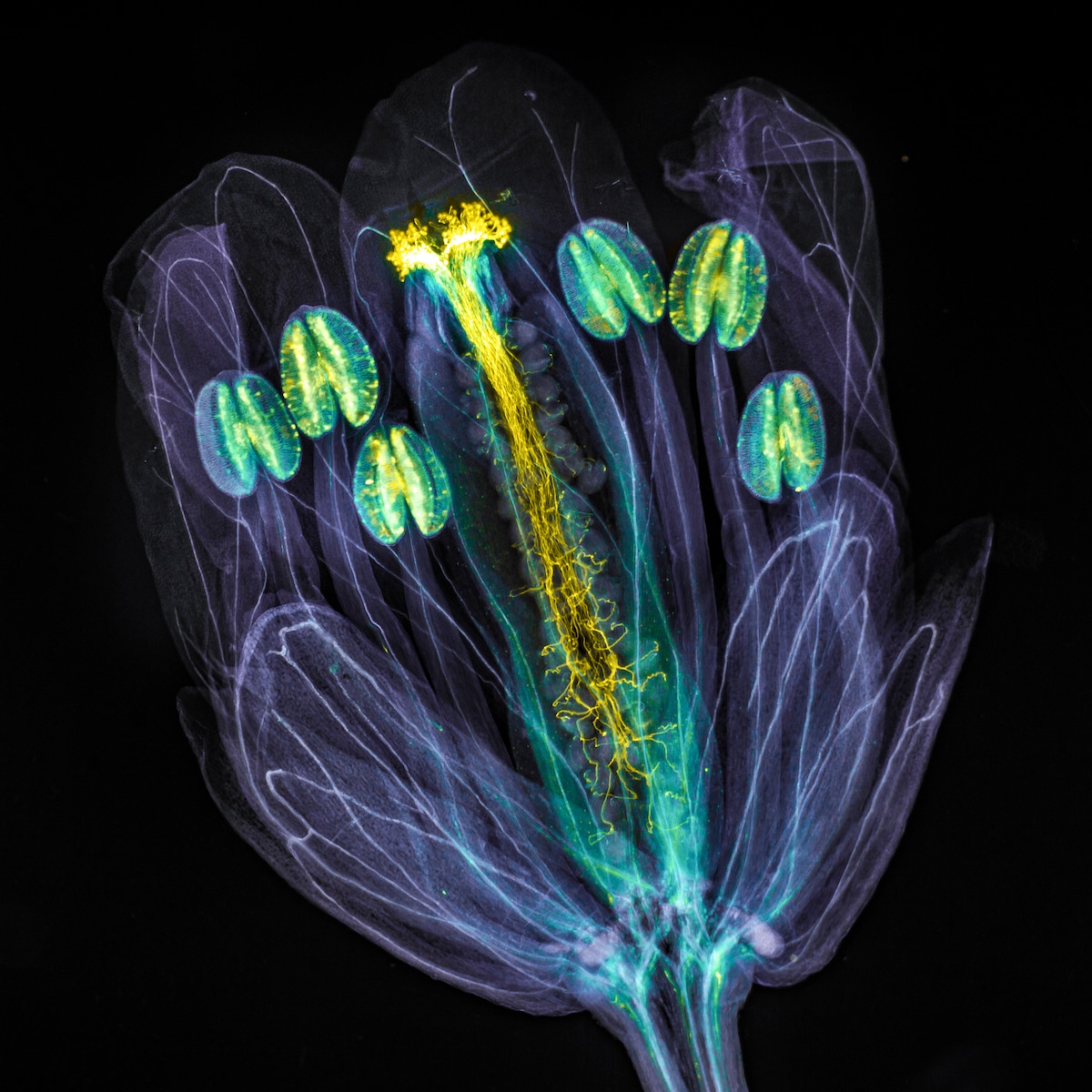
Global winner – Jan Martinek (Czech Republic).
“Arabidopsis thaliana flower with pollen tubes growing through the pistil. The flower tissues were chemically cleared to become transparent, while the pollen tubes were stained with aniline blue (yellow fluorescence) in order to be seen.”
Nearly 800 images were submitted to the Olympus Image of the Year Award 2021. Celebrating scientific photos taken with a microscope, the contest is filled with imagery that reveals a world hidden from the naked eye. This year’s winner, Jan Martinek, took the top prize thanks to his interesting look at a small flowering plant often used in genetic research.
Martinek, who hails from the Czech Republic, is a plant cell biologist and spends many hours looking at plants under a microscope. He takes hundreds of images, which he uses in his research and that he also publishes on social media for the enjoyment of the scientific community. “I am happy that thanks to the IOTY Award, my and others’ pictures of this fascinating microcosm get worldwide attention,” he shared.
The contest is run by Evident Life Science, formerly Olympus Scientific Solutions, which had an expert carefully pore over the images to find those that marry technical skills and artistry. Aside from the grand prize, regional winners were also recognized, and with photographers from 49 countries, there was plenty of global talent to reward. From a unique look at moss to the ovaries of a fruit fly, the subjects of the photographs demonstrate the artistic wonders hidden inside ordinary items.
“I continue to be amazed by the creative images we see each year that turn life under the microscope into unique art pieces,” said Satoshi Nakamura, vice president of Scientific Solutions Global Marketing at Evident. “We are honored to receive so many stunning captures from around the world that visually blend artistic creativity and science. The winning images create a beautiful image gallery showcasing an incredible level of talent and technique at the microscope.”
All of the winners received Olympus microscopes to further their scientific and artistic pursuits. If you can’t get enough of microscopy, check out the contest page for last year’s winners, which have even been transformed into virtual backgrounds for your next online meeting.
Olympus Image of the Year Award 2021 celebrates the best microscopy across the globe.
Regional Winner (Americas) – Ivan Radin (USA).
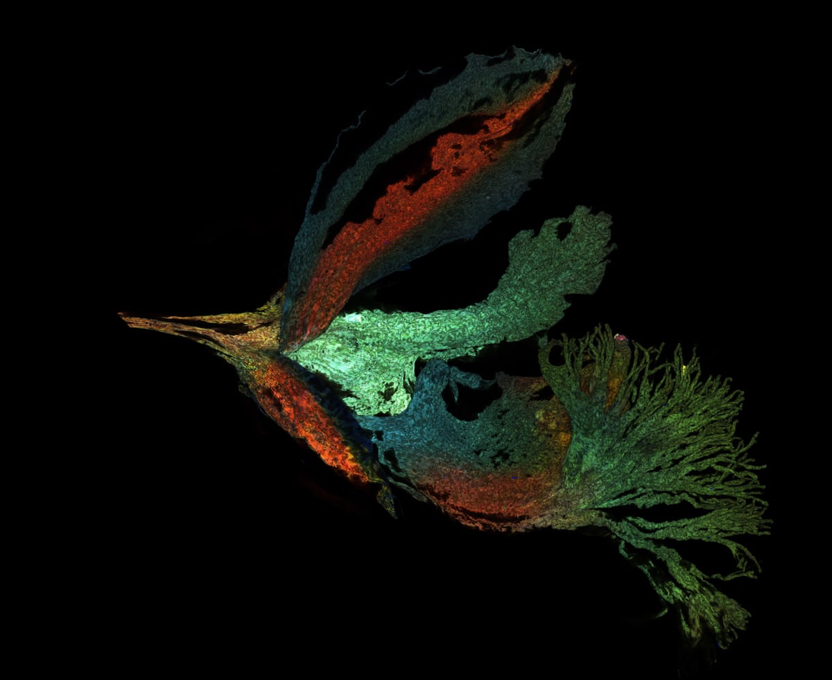
“A maximum projection of the deconvolved Z-stack of moss Physcomitrium patens protonemal cells. Cell walls (in cyan) were stained live with calcofluor white. Chloroplasts autofluorescence is in Fall LUT.”
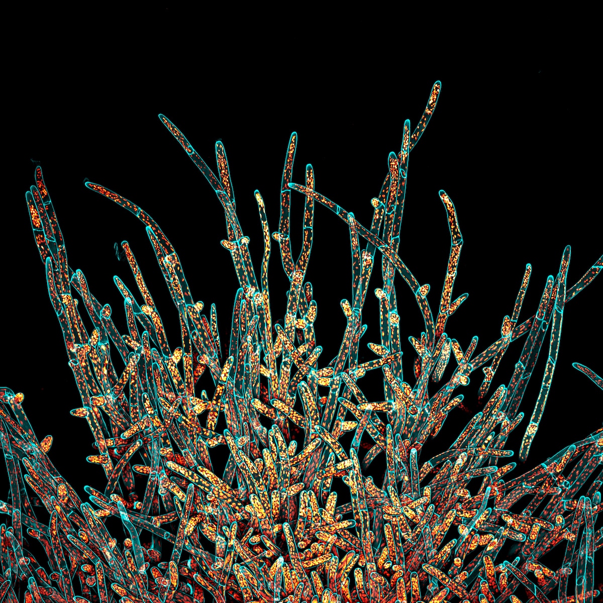
Regional winner (Asia-Pacific) – Daniel Han (Australia).
“Fern sori capsules with spores bursting out. Captured using Z-stacking.”
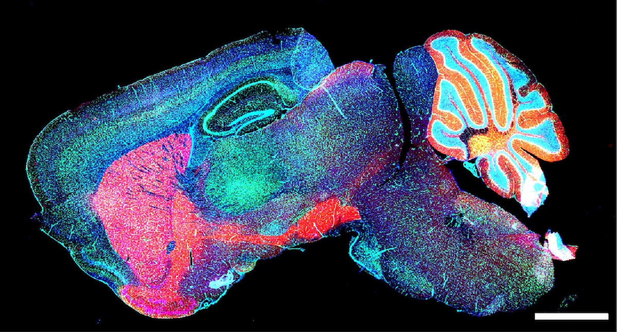
Honorable mention – Yayun Wang (China).
“Mouse brain GABA neurons.”
Over 800 images taken with microscopes from 49 countries were submitted to the contest.
Honorable mention – Mingyue Jia (China).
“Autofluorescence image of Siberian polygala. Captured using confocal microscopy. Rendered using maximum projection.”
Honorable mention – Yujun Chen (USA).
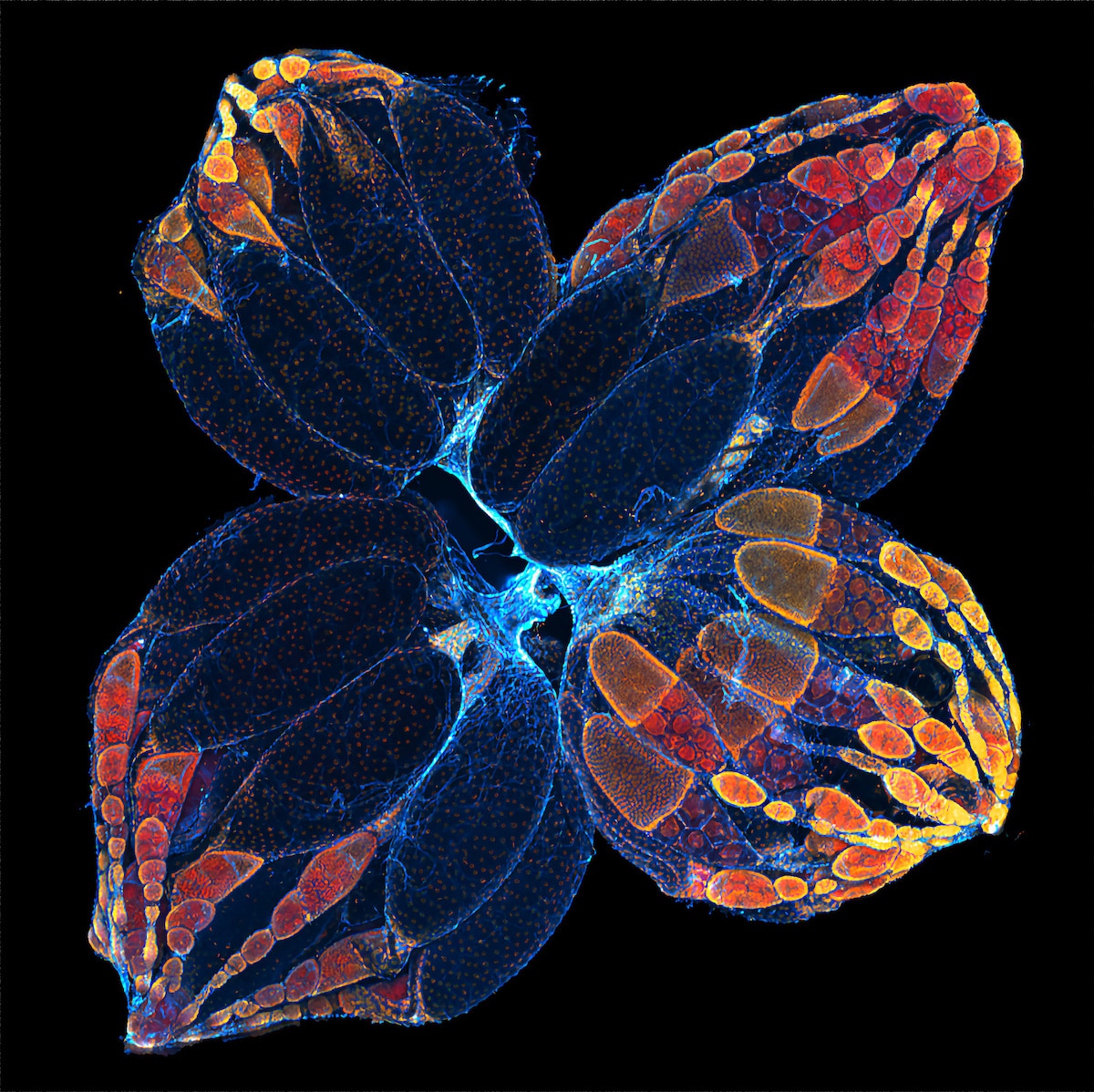
“Ovaries of the fruit fly.”
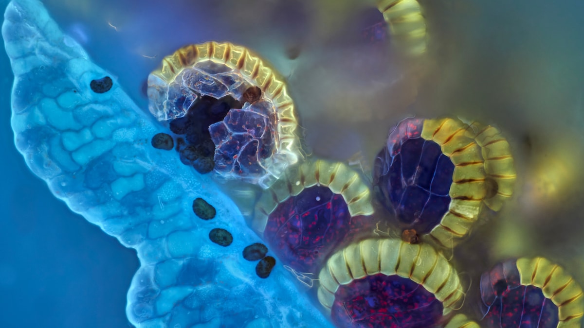
Regional winner (Europe, the Middle East, and Africa) – Vasilis Kokkoris (Netherlands).
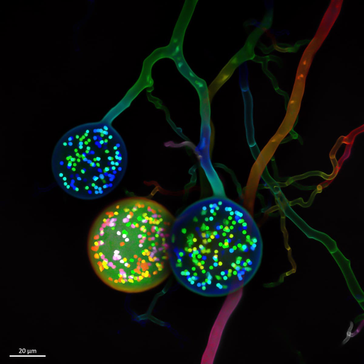
“Multinucleate spores of a soil fungus. One cell typically carries one nucleus. In contrast, as seen here, an arbuscular mycorrhizal (AM) fungal cell carries hundreds of nuclei.”
Honorable mention – Igor Siwanowiczfrom (USA).
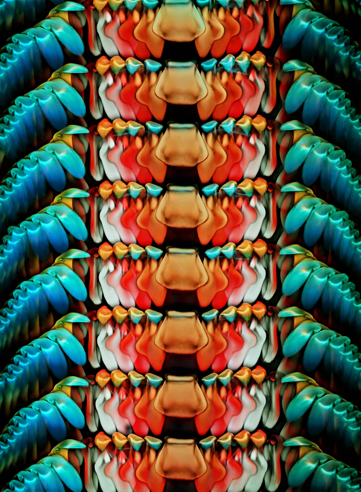
“Rasping tongue, or radula, of an Astraea conehead snail, Astraea tecta. Stained with Congo red. Imaged using a 10X (0.45 NA) objective. Depth color-coded projection.”
Overall and regional winners were awarded and several images were highlighted as honorable mentions.
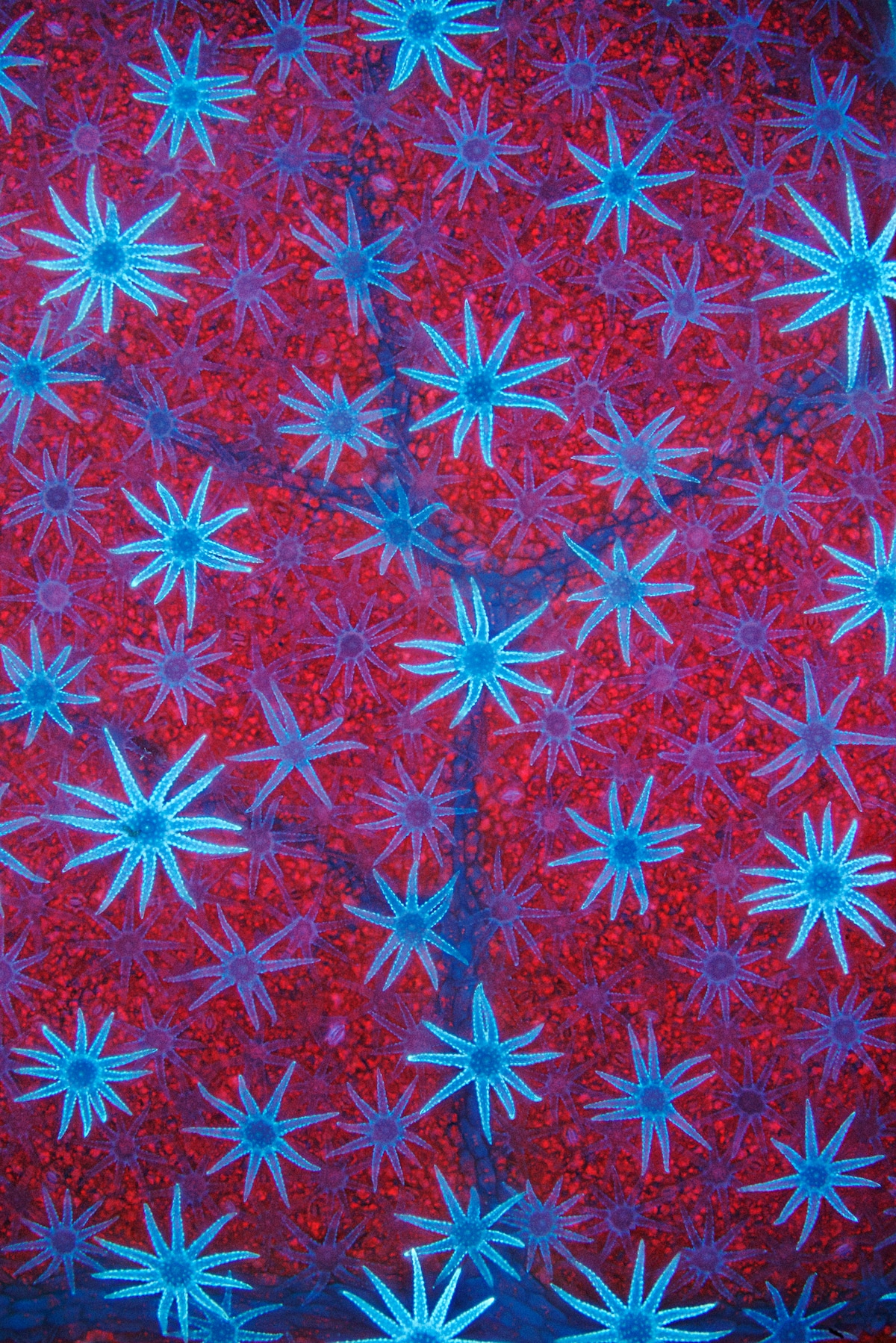
Honorable mention – David Maitland (UK).
“Blue autofluorescent, star-shaped defensive hairs cover the surface of a Deutzia leaf. The hairs are silhouetted against the leaf’s red-fluorescent, chlorophyll-packed cells.”
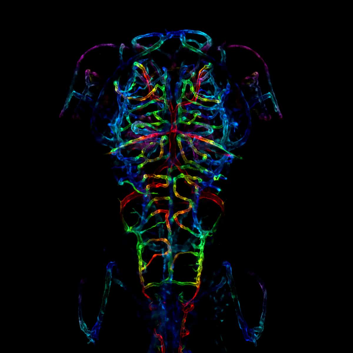
Honorable mention – LayraCintron-Rivera (USA).
“The developing nervous system of an embryonic zebrafish. Specifically, the image is a color-coded projection of the axonal projections of a zebrafish fixed six days after fertilization.”
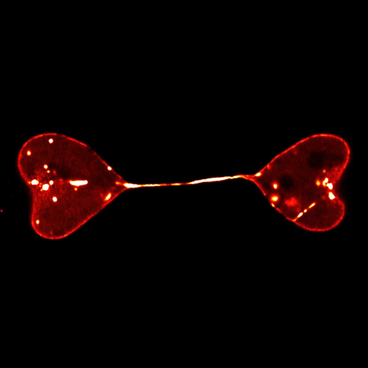
Honorable mention – Di Lu (China).
“Semi-separated nuclei of two cells form a heart-to-heart shape. The nuclei were labeled by lamin.”